
MRI, CT Scan, X-Ray and Ultrasound: Understanding Diagnostic Imaging
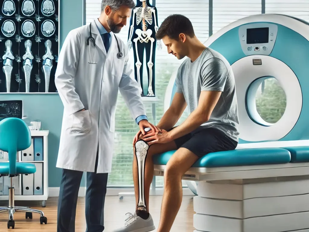
Accurate diagnosis is one of the most important steps in effective medical treatment. Before a doctor recommends medication, surgery, or another treatment, it is often necessary to understand what is happening inside the body. This is where diagnostic imaging plays an important role.
Modern imaging technologies allow doctors to examine bones, organs, blood vessels, soft tissues, and other structures without always requiring invasive procedures. Among the most commonly used imaging techniques are MRI, CT Scan, X-Ray, and Ultrasound. Each technology works differently and provides different types of information.
Choosing the right imaging test depends on factors such as the patient’s symptoms, suspected condition, body part being examined, urgency of the situation, age, medical history, and whether contrast material is required.
At Delta Hospitals, Radiology services support the diagnosis and management of a wide range of medical conditions. Imaging may be used for emergency care, routine evaluation, surgical planning, cancer assessment, orthopaedic conditions, neurological disorders, abdominal problems, and many other clinical situations.
For patients looking for diagnostic imaging in Rajahmundry, understanding the difference between MRI, CT Scan, X-Ray, and Ultrasound can make medical investigations easier to understand and less confusing.
What Is Diagnostic Imaging?
Diagnostic imaging refers to medical technologies used to create images of structures inside the body.
Imaging can help doctors:
- Identify injuries
- Detect diseases
- Locate tumors
- Examine organs
- Assess fractures
- Evaluate blood vessels
- Plan surgery
- Monitor treatment
No single imaging test is best for every condition. The doctor selects the most appropriate test based on the clinical situation.
X-Ray: A Fast and Common Imaging Test
An X-Ray is one of the oldest and most widely used forms of medical imaging. It uses a small amount of ionizing radiation to create images of internal structures.
X-rays are particularly useful for evaluating bones and certain chest conditions.
Common Uses of X-Ray
X-rays may help diagnose:
- Bone fractures
- Joint injuries
- Arthritis-related changes
- Chest infections
- Certain lung abnormalities
- Pneumonia
- Spinal alignment problems
A chest X-ray may be recommended when a patient has persistent respiratory symptoms or certain chest-related concerns.
Benefits of X-Ray
X-Ray imaging offers several advantages:
- Quick examination
- Widely available
- Useful for emergency situations
- Effective for detecting many fractures
- Relatively simple procedure
Because X-rays are quick, they are frequently used in Emergency Medicine when doctors need to assess injuries rapidly.
At Delta Hospitals, X-Ray imaging can support emergency, orthopaedic, pulmonary, and general medical evaluation.
Limitations of X-Ray
Although useful, X-rays do not provide the same level of soft-tissue detail as MRI.
They may not clearly show:
- Muscles
- Ligaments
- Tendons
- Certain internal organs
- Detailed brain structures
When more detailed imaging is required, the doctor may recommend CT, MRI, or Ultrasound.
CT Scan: Detailed Cross-Sectional Imaging
A CT Scan, or Computed Tomography scan, combines multiple X-ray images with computer processing to produce detailed cross-sectional images of the body.
Unlike a conventional X-ray, which produces a relatively flat image, CT creates detailed slices that allow doctors to examine internal structures from multiple views.
When Is a CT Scan Used?
CT imaging is particularly useful for:
- Head injuries
- Brain bleeding
- Severe trauma
- Abdominal pain
- Kidney stones
- Lung conditions
- Tumors
- Internal bleeding
- Complex fractures
- Certain vascular conditions
CT is especially valuable in emergency medicine because the examination can be performed relatively quickly.
CT Scan in Emergency Care
When a patient arrives after a serious accident, doctors may need to determine whether there is:
- Internal bleeding
- Brain injury
- Organ damage
- Complex fractures
CT imaging can provide detailed information rapidly, helping doctors make important treatment decisions.
This is one reason advanced CT Scan services in Rajahmundry are important for hospitals providing emergency and trauma care.
Advantages of CT Scan
Important benefits include:
- Detailed images
- Rapid imaging
- Excellent emergency assessment
- Useful bone and organ visualization
- Effective evaluation of trauma
CT can provide information that may not be visible on a conventional X-ray.
Limitations of CT Scan
CT uses ionizing radiation, so doctors recommend it when the expected diagnostic benefit justifies the exposure.
Some CT studies may also require contrast material.
Patients should inform their healthcare team about relevant allergies, kidney problems, pregnancy, or other medical conditions before a contrast-enhanced CT examination.
MRI: Detailed Soft-Tissue Imaging
Magnetic Resonance Imaging (MRI) uses powerful magnetic fields and radio waves to create detailed images of the body.
Unlike X-rays and CT scans, MRI does not use ionizing radiation.
MRI is particularly useful for viewing:
- Brain
- Spinal cord
- Spine
- Muscles
- Ligaments
- Tendons
- Joints
- Other soft tissues
Common Uses of MRI
Doctors may recommend MRI for:
- Brain disorders
- Stroke assessment in selected situations
- Brain tumors
- Spine problems
- Disc disorders
- Joint injuries
- Ligament tears
- Muscle injuries
- Certain cancers
- Soft-tissue abnormalities
MRI can provide detailed images that are especially useful when soft-tissue structures need to be evaluated.
MRI for Brain and Spine Conditions
MRI is frequently used in neurological and spine evaluation because it provides detailed images of the brain, spinal cord, discs, and surrounding tissues.
It can help doctors evaluate conditions such as:
- Disc prolapse
- Spinal stenosis
- Brain tumors
- Certain vascular abnormalities
- Nerve compression
- Other neurological disorders
At Delta Hospitals, MRI imaging can support Neurology, Neurosurgery, Orthopaedics, and other specialist departments when clinically indicated.
Advantages of MRI
Key advantages include:
- Excellent soft-tissue detail
- No ionizing radiation
- Detailed brain and spine imaging
- Useful for muscles, ligaments, and joints
MRI is particularly valuable when an X-ray or CT scan does not provide enough information.
MRI Limitations
MRI examinations may take longer than an X-ray or some CT scans.
Some patients may also find the enclosed scanner uncomfortable.
People with certain implanted medical devices or metal fragments may require special evaluation before undergoing MRI.
Patients should provide complete information about implants, previous surgeries, and other relevant medical history.
Ultrasound: Safe and Real-Time Imaging
Ultrasound uses high-frequency sound waves to create images of structures inside the body.
It does not use ionizing radiation and can provide real-time imaging.
Ultrasound is commonly used to examine:
- Liver
- Gallbladder
- Kidneys
- Thyroid
- Pelvic organs
- Blood vessels
- Soft tissues
It is also widely used during pregnancy.
Common Uses of Ultrasound
Ultrasound may help identify:
- Gallstones
- Kidney stones
- Cysts
- Abnormal fluid collections
- Organ abnormalities
- Thyroid nodules
- Certain blood-flow problems
It can also be used to guide certain procedures when appropriate.
Benefits of Ultrasound
Important advantages include:
- No ionizing radiation
- Real-time imaging
- Non-invasive
- Useful for many organs
- Can be performed repeatedly when clinically appropriate
Because of these advantages, ultrasound is an important part of modern diagnostic imaging.
MRI vs CT Scan
Patients often ask whether an MRI or CT Scan is better. The truth is that they are designed for different purposes.
MRI
Generally preferred when doctors need detailed images of:
- Brain
- Spine
- Soft tissues
- Ligaments
- Tendons
- Certain joints
CT Scan
Often preferred when doctors need:
- Rapid emergency imaging
- Detailed bone assessment
- Trauma evaluation
- Detection of internal bleeding
- Detailed abdominal or chest imaging
The doctor chooses the test based on the suspected condition.
CT Scan vs X-Ray
Both CT and X-Ray use X-rays, but they provide different levels of detail.
X-Ray
Best suited for many:
- Fractures
- Basic chest evaluations
- Bone assessments
CT Scan
Provides more detailed cross-sectional images and is particularly valuable for:
- Complex trauma
- Internal bleeding
- Tumors
- Organ abnormalities
An X-ray may be enough for a simple problem, while a CT may be needed when more detailed information is required.
Ultrasound vs MRI
Ultrasound and MRI are both useful for soft-tissue evaluation, but they have different roles.
Ultrasound
- Real-time imaging
- No radiation
- Useful for abdominal and superficial structures
- More accessible for certain examinations
MRI
- Highly detailed soft-tissue imaging
- Useful for brain and spine
- Useful for joints and complex soft tissues
The choice depends on the clinical question the doctor needs to answer.
Role of Contrast in Diagnostic Imaging
Some imaging examinations use contrast material to make specific structures easier to see.
Contrast may help doctors evaluate:
- Blood vessels
- Tumors
- Inflammation
- Organ structures
Not every scan requires contrast.
Before contrast-enhanced imaging, patients should inform their doctor about:
- Previous contrast reactions
- Kidney problems
- Pregnancy
- Relevant medications
- Other medical conditions
The healthcare team decides whether contrast is appropriate.
Diagnostic Imaging in Emergency Medicine
Diagnostic imaging is particularly important during medical emergencies.
Patients arriving with serious injuries may require rapid imaging to identify:
- Fractures
- Brain injuries
- Internal bleeding
- Organ damage
Patients with sudden neurological symptoms may require urgent imaging to determine the cause.
At Delta Hospitals, Radiology works alongside Emergency Medicine and Critical Care when rapid diagnosis is required.
Imaging for Cancer Diagnosis and Monitoring
Diagnostic imaging plays a major role in cancer care.
Depending on the cancer, imaging may help:
- Detect suspicious masses
- Determine tumor location
- Assess disease spread
- Support biopsy planning
- Monitor treatment response
MRI, CT, Ultrasound, and X-Ray each have different roles in cancer evaluation.
At Delta Hospitals, Radiology works with Medical Oncology, Surgical Oncology, and Radiation Oncology as part of multidisciplinary cancer care.
Imaging for Orthopaedic Conditions
Bone and joint problems often require imaging to determine the cause of pain or injury.
X-Ray
Useful for:
- Fractures
- Arthritis
- Bone alignment
CT Scan
Useful for:
- Complex fractures
- Detailed bone anatomy
MRI
Useful for:
- Ligament injuries
- Meniscus problems
- Disc disorders
- Tendon injuries
- Soft-tissue conditions
The appropriate test depends on the suspected injury or disease.
How to Prepare for Diagnostic Imaging
Preparation depends on the specific examination.
Some tests may require:
- Fasting
- Drinking water
- A full bladder
- Removal of metal objects
- Changing into hospital clothing
The hospital will provide specific instructions before the procedure.
Patients should inform the healthcare team about:
- Pregnancy
- Allergies
- Kidney disease
- Previous surgeries
- Implanted devices
- Current medications
Following preparation instructions can help ensure high-quality images and safe testing.
Is Diagnostic Imaging Safe?
The safety of imaging depends on the technology and the patient’s individual circumstances.
X-rays and CT scans use ionizing radiation, while MRI uses magnetic fields and radio waves, and ultrasound uses sound waves.
Doctors carefully consider the need for each examination and select the appropriate test based on the medical situation.
Patients should follow their doctor’s recommendations rather than avoiding an appropriate test because of general concerns about imaging.
Why Choose Delta Hospitals for Diagnostic Imaging?
Delta Hospitals provides access to modern diagnostic services that support multiple medical departments.
Our diagnostic capabilities include:
- MRI
- CT Scan
- Digital X-Ray
- Ultrasound
- Laboratory investigations
- Cardiac diagnostics
- Interventional Radiology
These services support:
- General Medicine
- Cardiology
- Neurology
- Neurosurgery
- Orthopaedics
- Pulmonology
- Urology
- Surgical Oncology
- Medical Oncology
- Emergency Medicine
This multidisciplinary environment helps patients receive appropriate investigations and coordinated medical care.
Choosing the Right Imaging Test
There is no universal “best” imaging test.
The correct examination depends on:
- Symptoms
- Body part being assessed
- Suspected disease
- Urgency
- Age
- Medical history
- Previous imaging
- Need for contrast
For example, a patient with a suspected simple fracture may only need an X-ray, while someone with a complex spinal problem may require an MRI.
A patient involved in major trauma may need CT because rapid, detailed imaging is important.
A child or pregnant patient may be evaluated with ultrasound when appropriate because it does not use ionizing radiation.
The Importance of Accurate Diagnostic Imaging
MRI, CT Scan, X-Ray, and Ultrasound each serve different diagnostic purposes. X-Ray is useful for many bone and chest conditions, CT provides detailed imaging for emergencies and internal injuries, MRI offers excellent soft-tissue images, and Ultrasound provides real-time imaging without radiation.
At Delta Hospitals, advanced Radiology services support diagnosis across multiple specialties. The appropriate imaging test depends on your symptoms, condition, and doctor’s assessment.
FAQs
1 . What is diagnostic imaging?
It uses imaging tests to identify diseases, injuries, and internal abnormalities.
2 . What is an X-Ray used for?
It is commonly used to examine bones, fractures, and chest conditions.
3 . What is a CT Scan used for?
CT provides detailed images for trauma, internal organs, tumors, and emergencies.
4 . What is an MRI used for?
MRI provides detailed images of the brain, spine, joints, and soft tissues.
5 . What is Ultrasound used for?
It provides real-time images of organs and soft tissues without radiation.
6 . Which is better, MRI or CT Scan?
Neither is universally better; the appropriate test depends on the condition.
7 . Do diagnostic imaging tests use radiation?
X-Ray and CT use radiation, while MRI and Ultrasound do not use ionizing radiation.
8 . Are diagnostic imaging tests safe?
They are generally safe when recommended and performed appropriately.
9 . Does Delta Hospitals provide diagnostic imaging services?
Yes, Delta Hospitals provides MRI, CT Scan, X-Ray, Ultrasound, and other Radiology services.
10 . How is the right imaging test selected?
A doctor selects the test based on symptoms, medical history, and suspected condition.